Scaphoid Fracture of the Wrist
|
||||||


Introduction
Physiotherapy in Edmonton for Wrist
Welcome to In Step Physical Therapy's patient resource about Scaphoid Fracture of the Wrist.
Doctors commonly diagnose a sprained wrist after a patient falls on an outstretched hand. However, if pain and swelling don't go away, doctors become suspicious that the injury is actually more serious. A fall on an outstretched hand commonly breaks the scaphoid bone of the wrist. X-rays taken at the time of the injury may not clearly show the fracture. If the fracture is not recognized early, it may not heal properly. This can lead to problems later.
This guide will help you understand:
- what causes fractures of the scaphoid bone
- what nonunion of the scaphoid bone is
- what you can do to treat each condition
#testimonialslist|kind:all|display:slider|orderby:type|filter_utags_names:Hand Pain|limit:15|heading:Hear from some of our patients who we treated for *Hand Pain*#
Anatomy
Where is the scaphoid bone of the wrist?
The anatomy of the wrist joint is extremely complex, probably the most complex of all the joints in the body. The joint is actually a collection of many joints and many bones. These joints and bones let us use our hands in many ways. The wrist must be extremely mobile to give our hands a full range of motion. At the same time, the wrist must provide the strength for heavy gripping.
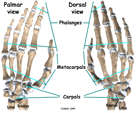
The wrist is made up of eight separate small bones, called the:
Carpal Bones
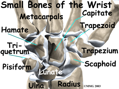
The scaphoid bone is a carpal bone near the base of the thumb. The carpal bones connect the two bones of the forearm, the radius and the ulna, to the bones of the hand. The metacarpal bones are the long bones that lie underneath the palm.
 The metacarpals attach to the phalanges, which are the bones in the fingers and thumb.
One reason that the wrist is so complicated is because every small bone forms a joint with the bone next to it.
This means that what we call the wrist joint is actually made up of many small joints. Ligaments connect all the small bones to each other, and to the radius, ulna, and metacarpal bones.
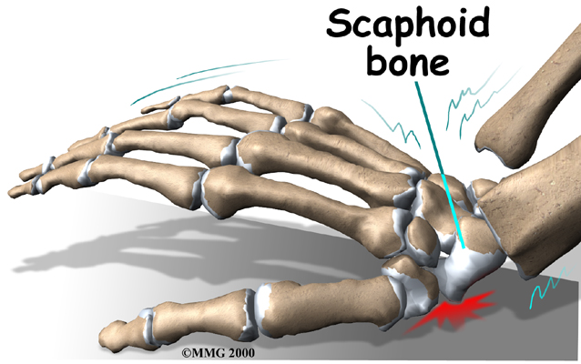
The scaphoid bone is a small carpal bone on the thumb side (radial side) of the wrist. It is the most commonly fractured carpal bone.
This is probably because it actually crosses two rows of carpal bones, forming a hinge.
A fall on the outstretched hand puts heavy stress on the scaphoid bone. This stress can cause either a small crack through the middle of the bone or a complete separation of the bone into two pieces. A separation is called a displaced fracture.
Related Document: In Step Physical Therapy's Guide to Wrist Anatomy
Causes
 What causes a scaphoid fracture?
A scaphoid fracture is almost always caused by a fall on the outstretched hand. We commonly try to break a fall by putting our hands out for protection. Landing on an outstretched hand makes hand and wrist injuries, including a fracture of the scaphoid bone, fairly common.
When a scaphoid fracture is recognized on the first X-ray, treatment begins immediately. But patients often assume that the injury is just a sprain, and they wait for it to heal on its own. In some cases, the wrist gets better. In many cases the bone fails to heal. The scaphoid fracture then develops into what surgeons call a nonunion.
 A nonunion can occur in two ways. In a simple nonunion, the two pieces of bone fail to heal together.
The second type of nonunion is much more serious.
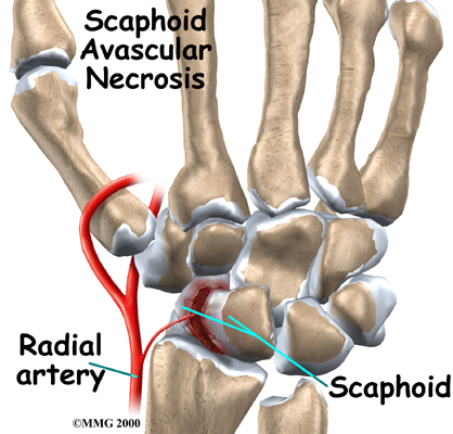
The lower half of the fractured bone loses its blood supply and actually dies.
This condition is called avascular necrosis (Avascular means no blood supply, and necrosis means dead.)
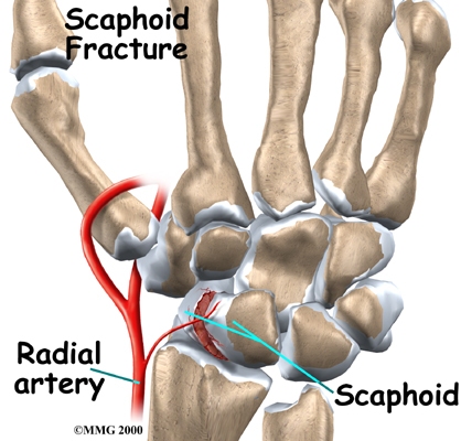
The scaphoid bone is at risk for avascular necrosis.
Only one small artery enters the bone, at the end that is closest to the thumb.
If the fracture tears the artery, the blood supply is lost. Avascular necrosis becomes easy to see on X-rays several months after the injury.
Symptoms
How will I know if I have a scaphoid fracture?
The symptoms of a fresh fracture of the scaphoid bone usually include pain in the wrist and tenderness in the area just below the thumb. You may also see swelling around the wrist. The swelling occurs because blood from the fractured bone fills the wrist joint. Thin people will see a bulging of the joint capsule. The joint capsule is the watertight sac that encloses the joint.
Symptoms of a nonunion of the scaphoid bone are more subtle. You may have pain when you use your wrist. However, the pain may be very minimal. It is fairly common for doctors to see a nonunion of the scaphoid bone on X-rays, but the patient can't remember an injury. These people probably suffered a wrist injury years ago that they thought was a simple sprain. Still, the most common symptom of a nonunion is a gradual increase in pain. Over several years the nonunion can lead to degenerative arthritis in the wrist joint.
Diagnosis
How will my health care provider identify the problem?
When you visit In Step Physical Therapy, our physiotherapist will first take a medical history. We will ask you questions about your pain and about any injuries to your wrist. Our therapist will also do a physical exam. The prodding and moving may hurt your wrist a bit. But it is important that your doctor know exactly where your pain is coming from.
It is usually safe to assume that any patient who has fallen on an outstretched hand and has swelling or tenderness on the thumb side of the wrist has a scaphoid fracture. You should assume this until tests prove otherwise.
Some patients may be referred to a doctor for further diagnosis. Once your diagnostic examination is complete, the physiotherapists at In Step Physical Therapy have treatment options that will help speed your recovery, so that you can more quickly return to your active lifestyle.
Our Treatment
Non-surgical Rehabilitation
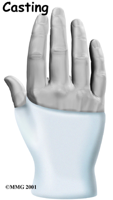
If the bone is in good alignment, and there are no problems with the blood supply to the bone, you may be placed in a cast for nine to 12 weeks. Some doctors prefer to start with a long-arm cast. Others use a thumb-spica cast designed to keep the wrist and thumb from moving.
The amount of time you need to wear the cast depends on what part is fractured and whether the bones heal well. When your doctor is certain the bones have healed, your cast will be removed. Your wrist will probably be stiff and weak from being in the cast. You can then begin your In Step Physical Therapy rehabilitation program to help improve wrist range of motion and strength.
Post-surgical Rehabilitation
Depending on the type of surgery you have, you may be placed in a splint for up to 12 weeks after surgery. Your surgeon will X-ray the wrist several times after surgery to make sure that the bones are healing properly. Once the two halves of the scaphoid bone have healed, you can safely begin your In Step Physical Therapy rehabilitation program.
Our first few physiotherapy treatments will focus on controlling the pain and swelling. Our physiotherapist will gradually have you work into doing exercises that will help strengthen and stabilize the muscles around the wrist joint. We use other exercises to improve fine motor control and dexterity of your hand. Our physiotherapist will give you tips on ways to do your activities while avoiding extra strain on the wrist joint.
In Step Physical Therapy provides services for physiotherapy in Edmonton.
Physician Review
 X-rays taken immediately after the injury may not show a fracture. Still, most surgeons will put a cast on the wrist and get another X-ray in 10 days. This gives the edges of the fractured bone time to heal, and may prevent nonunion. By waiting 10 days, the fracture is easier to see on an X-ray.
If it is still not clear whether or not you have a fracture, your doctor may order other imaging tests. You may have a bone scan done. A bone scan involves injecting tracers into your blood stream. The tracers then show up on special X-rays of your wrist. The tracers build up in areas of extra stress to bone tissue, such as a fracture.
Your doctor may also order a magnetic resonance imaging (MRI) scan. An MRI scan is a special imaging test that uses magnetic waves to create pictures of your body in slices.
The MRI scan shows tendons as well as bones. It is painless and requires no needles or injections.
Fracture
If the fracture is identified immediately and is in good alignment, you will probably wear a cast for nine to 12 weeks. The cast will cover your forearm, wrist, and thumb. This is necessary to hold the scaphoid bone very still while it heals.
Your doctor will take X-rays at least once a month to check the progress of the healing. Once your doctor is sure the fracture has healed, the cast will be removed. Even with this type of treatment, there is still a risk that the fracture may not heal well and will become a nonunion.
Nonunion
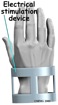
A fracture that doesn't heal within several months is considered a nonunion. If the injury is fairly recent, your doctor might recommend more time in the cast. He or she might also prescribe an electrical stimulator. The electrical stimulator is a device that sends a small electrical current to your scaphoid bone. You wear it like a large bracelet for 10 to 12 hours a day. Electrical current has been shown to help the bones heal.
Surgery
Screw Fixation
Some surgeons report good results doing surgery right away when a patient has had a recent, nondisplaced scaphoid fracture. Studies have shown that this method can help people get back to activity faster than wearing a cast for up to 12 weeks. The procedure involves inserting a screw through the scaphoid. The screw holds the scaphoid firmly until it heals.
Scaphoid Debridement
In cases where a nonunion has occurred depite wearing a cast and using an electrical stimulator, surgery will likely be suggested. An incision is made in the wrist directly over the scaphoid bone. The surgeon finds the old fracture line on the scaphoid bone. All the scar tissue between the two halves of the bone must be removed (debrided). This creates a fresh bone surface to allow healing to begin again. In some cases, damaged bone tissue from the scaphoid is also removed.
Bone Graft Method
Your surgeon may use a bone graft. A bone graft involves taking bone tissue from another spot in your wrist and inserting it into the fracture. A bone graft can stimulate healing on the surface of the bones. The bone graft is usually taken through a second small incision just above the wrist. (It is sometimes taken from the pelvis, through an incision in the side of your hip.)
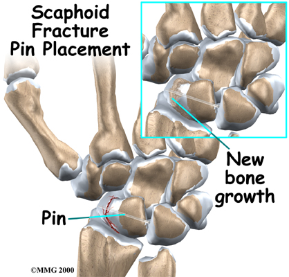
After the bone graft is placed between the parts of the scaphoid bone, some surgeons also insert a metal pin or screw across the bone. The goal is to hold the two pieces of bone tightly together, allowing them to fuse into one bone.
When the surgery is complete, the incision is stitched closed. The arm is placed in a large bandage or a splint. You are then awakened and taken to the recovery room.
Sometimes the bones still do not heal as planned. Surgeons call a fused bone that fails to heal a pseudarthrosis. If the nonunion continues to cause pain, you may need a second operation. Your surgeon will probably add more bone graft and check that the pins or screws are holding the bones together.
Portions of this document copyright MMG, LLC.
